The Importance of Histopathology Biopsy in Accurate Cancer Diagnosis
What is the purpose of a histopathology biopsy?
Histopathology biopsy is a medical procedure that involves the microscopic examination of tissues or cells to diagnose a disease or condition. The primary purpose of a histopathology biopsy is to obtain a tissue sample from a patient and analyze it under a microscope to identify any abnormalities or changes in the tissue structure or function.
A histopathology biopsy test is crucial in the diagnosis, treatment, and management of various medical conditions such as cancer, infections, inflammatory diseases, and autoimmune disorders. The biopsy provides information about the type, stage, and aggressiveness of the disease, which helps doctors create an effective treatment plan for the patient.
The biopsy procedure involves the removal of a small piece of tissue or cells from the affected area using a special instrument, such as a needle, scalpel, or punch. The tissue sample is then sent to a laboratory for analysis by a qualified pathologist who examines it under a microscope and produces a report on the findings.
Histopathology biopsy plays a critical role in oncology. It provides important information about the tumor's size, grade, and location, which helps oncologists determine the best course of treatment, such as surgery, chemotherapy, radiation therapy, or immune therapy.
In summary, histopathology biopsy is a vital medical procedure that enables doctors to diagnose diseases accurately, plan and provide appropriate treatment, and monitor disease progression. The procedure is safe, minimally invasive, and can provide life-saving information to patients and their caregivers.
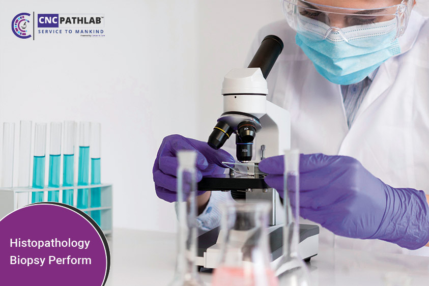
How is a histopathology biopsy performed?
Histopathology biopsy is a medical procedure that involves the surgical removal of a small piece of tissue from a patient's body for further laboratory analysis. This procedure is commonly used to diagnose various medical conditions and diseases, including cancer. Here is a step-by-step guide on how a histopathology biopsy is performed:
Step 1: Preparation
Before the procedure, the patient is usually given a local anesthetic to numb the area where the biopsy will be performed. The healthcare provider will then clean the area around the biopsy site with an antiseptic solution to prevent infection.
Step 2: Tissue removal
A small incision is made in the skin to access the affected area, and a special tool called a biopsy needle is inserted through the incision to remove a small piece of tissue. In some cases, a larger surgical instrument may be used to remove a larger tissue sample.
Step 3: Tissue handling
Once the tissue sample has been removed, it is immediately placed in a container filled with a special preservative solution to prevent decay and damage. The container is then transported to a laboratory for further analysis.
Step 4: Laboratory analysis
In the laboratory, the tissue sample is prepared for analysis by a histotechnologist, who slices the tissue into thin sections and stains them with special dyes to highlight different structures within the cells. The sample is then examined under a microscope by a pathologist, who will interpret the results and make a diagnosis.
Step 5: Results
Once the histopathology biopsy results are available, the pathologist will communicate them to the patient's healthcare provider, who will then discuss them with the patient and their caregiver. The results may show a diagnosis of a particular medical condition or disease, such as cancer. The results may also help the healthcare provider plan and provide appropriate treatment, such as surgery, chemotherapy, radiation therapy, or immune therapy. In some cases, the results may also help monitor disease progression and make necessary adjustments to the treatment plan.
In conclusion, histopathology biopsy is a crucial medical procedure that plays a significant role in diagnosing and treating various medical conditions and diseases, including cancer. Patients and their caregivers can rest assured that this procedure is safe, minimally invasive, and provides life-saving information that can help improve their quality of life. It is essential to consult with a healthcare provider to determine if a histopathology biopsy is necessary for accurate diagnosis and treatment planning.
What types of tissue samples are examined in a histopathology biopsy?
Histopathology biopsy is a diagnostic procedure that involves examining tissue samples under a microscope to identify any abnormal cellular changes or conditions. This procedure is crucial in diagnosing and treating various diseases, including cancer.
The types of tissue samples examined in a histopathology biopsy can vary depending on the suspected condition. However, some of the most common tissue samples examined include:
1. Skin Biopsy: This involves the removal of a small piece of skin tissue for examination. It is used to diagnose skin conditions such as psoriasis, eczema, and skin cancer.
2. Endoscopic Biopsy: This involves inserting a thin, flexible tube with a light and camera attached to it into the body to collect tissue samples from internal organs such as the stomach, lungs, and colon. This type of biopsy is used to diagnose conditions such as ulcers, inflammation, and cancer.
3. Needle Biopsy: This involves using a needle to collect a small amount of tissue from a tumor or abnormal growth. This type of biopsy is used to diagnose conditions such as breast cancer, lymphoma, and lung cancer.
4. Bone Marrow Biopsy: This involves collecting a small amount of bone marrow tissue from the hipbone to diagnose conditions such as leukemia, lymphoma, and multiple myeloma.
In conclusion, histopathology biopsy is a vital diagnostic tool that involves examining tissue samples under a microscope to identify any abnormal changes or conditions. The type of tissue sample collected depends on the suspected condition, and it is essential to consult with a healthcare provider to determine if this procedure is necessary for accurate diagnosis and treatment planning. The information gathered from a histopathology biopsy can provide life-saving information and improve the quality of life for patients and their caregivers. As healthcare professionals, it is our responsibility to educate and inform patients about the importance of this procedure and ensure they receive the best possible care. By working together, we can help diagnose and treat various medical conditions and diseases, including cancer, and improve the lives of those affected.
How is a histopathology biopsy sample prepared for analysis?
Histopathology biopsy samples are commonly used to diagnose various medical conditions. These samples are obtained from different parts of the body, such as the skin, liver, kidney, or lung. The preparation of a histopathology biopsy sample is crucial to ensure accurate analysis and diagnosis. Here are the steps involved in preparing a histopathology biopsy sample for analysis:
1. Fixation: The biopsy sample is first fixed in formalin, a solution that preserves the tissue's structure and prevents decomposition. The sample is usually immersed in formalin for 24-48 hours.
2. Dehydration: The sample is then dehydrated in a series of alcohol solutions, starting from low concentration to high concentration. This process removes water from the tissue and prepares it for embedding.
3. Embedding: The dehydrated tissue is embedded in paraffin wax, which provides support and makes it easier to cut thin sections. The paraffin blocks are then cooled and hardened.
4. Sectioning: The paraffin block is cut into thin sections (4-6 microns) using a microtome. The sections are then mounted on glass slides and dried.
5. Staining: The sections are stained with different dyes, depending on the type of tissue and the purpose of the analysis. Hematoxylin and eosin (H&E) stain is the most common stain used in histopathology, as it highlights the nucleus and cytoplasm of the cells and provides contrast between different tissues.
6. Examination: The stained sections are then examined under a microscope by a pathologist or trained specialist. They analyze the tissue's structure, cell type, and any abnormalities present, which helps to diagnose various medical conditions.
In conclusion, histopathology biopsy is a critical procedure that provides valuable information for accurate diagnosis and treatment planning. The preparation of the biopsy sample is a meticulous process that requires attention to detail and expertise. It involves fixation, dehydration, embedding, sectioning, staining, and examination under a microscope. By understanding the importance of this procedure and following the necessary steps, healthcare professionals can provide their patients with the best possible care and improve their quality of life.
What are the benefits of a histopathology biopsy?
Histopathology biopsy is a medical procedure that involves the examination and analysis of tissue samples to diagnose and treat various diseases, including cancer. This procedure is considered one of the most effective ways to diagnose and monitor various medical conditions, and it offers a wide range of benefits.
One of the primary benefits of histopathology biopsy is that it provides accurate and reliable results. The procedure involves the examination of tissue samples under a microscope, which allows medical professionals to identify abnormal cell growth, inflammation, infection, and other abnormalities that may be present. This level of detail and precision enables physicians to make informed decisions about treatment options and monitor the progression of the disease over time.
Another benefit of histopathology biopsy is that it is a minimally invasive procedure that can be performed on an outpatient basis. This means that patients can often return home the same day and resume their normal activities with minimal disruption. Additionally, the procedure is relatively painless and is usually performed under local anesthesia, which further reduces any discomfort.
Histopathology biopsy also allows for personalized treatment options. By accurately diagnosing the condition and identifying its specific characteristics, medical professionals can create a treatment plan that is tailored to the individual patient. This personalized approach can help improve the patient's outcomes and reduce the potential for complications or side effects.
Finally, histopathology biopsy can help prevent the spread of disease. By identifying abnormal tissue growth early, medical professionals can take steps to prevent the disease from spreading to other parts of the body. This can be especially important in the case of cancer, where early detection and treatment can significantly improve survival rates.
In conclusion, histopathology biopsy is an essential medical procedure that offers numerous benefits for patients. It provides accurate and reliable results, is minimally invasive, and allows for personalized treatment options. Ultimately, histopathology biopsy can help improve patient outcomes and prevent the spread of disease. If you are experiencing symptoms related to a medical condition or have concerns about your health, it is crucial to consult with a healthcare provider to determine if a histopathology biopsy may be necessary for accurate diagnosis and treatment planning. With the help of medical professionals and advanced diagnostic tools, you can take control of your health and enjoy a better quality of life.
How is a histopathology biopsy sample prepared for analysis?
Histopathology biopsy samples are critical tools used in the diagnosis of numerous medical conditions. These samples are typically collected during a biopsy procedure and must be carefully prepared for analysis in a laboratory setting.
The first step in the preparation of a histopathology biopsy sample is to fix it. This involves immersing the sample in a solution containing formaldehyde or another fixative. The purpose of this step is to preserve the tissue's cellular and architectural features, preventing them from being distorted or destroyed during subsequent processing.
After fixation, the sample is dehydrated using a series of solutions containing increasing concentrations of alcohol. This process helps remove water from the tissue, which could interfere with subsequent processing steps. The tissue is then cleared using a solution such as a xylene or another clearing agent, which helps to remove the alcohol.
Next, the sample is embedded in a solid medium such as paraffin wax, which provides support for the tissue during subsequent cutting and staining steps. Once the wax has solidified, the tissue block is cut into thin slices called sections using a microtome.
The sections are then mounted onto glass slides, and any remaining wax is removed using a clearing agent. The tissue sections are then stained using various methods, such as hematoxylin and eosin, which highlights different structures within the tissue.
Once stained, the slides are examined under a microscope by a trained pathologist or histotechnologist, who analyzes the tissue's cellular and architectural features to make a diagnosis.
In summary,a histopathology biopsy sample is carefully prepared for analysis to ensure accurate diagnosis and treatment planning. The process involves fixing the tissue, dehydrating it, clearing it, embedding it in wax, cutting it into sections, staining it, and examining it under a microscope. Each step must be performed with precision and attention to detail to ensure that the tissue's cellular and architectural features are preserved and accurately analyzed.
At the heart of this process are the skilled professionals who work tirelessly to ensure that each sample is analyzed accurately and efficiently. From the healthcare providers who collect the samples to the histotechnologists who prepare them for analysis, each member of the team plays a vital role in the diagnosis and treatment of medical conditions. By working together and leveraging the latest diagnostic tools and techniques, medical professionals can provide patients with the care and support they need to achieve optimal health and wellbeing.
Are there any special techniques used in a histopathology biopsy?
Histopathology is the study of tissue samples to identify diseases or abnormalities. Biopsy is the process of taking a small sample of tissue from a patient's body for laboratory analysis. In histopathology biopsy, there are specific techniques used to ensure that the sample is properly preserved and analyzed.
One of the essential techniques used in histopathology biopsy is fixation. Fixation is the process of preserving the tissue sample in formalin, which prevents the tissue from decomposing and helps to stabilize the sample. The sample is then embedded in paraffin wax, which helps to provide support and stability for the tissue during the cutting process.
Another important technique used in histopathology biopsy is staining. Staining is the process of coloring the tissue sample to highlight specific structures or cells. Hematoxylin and eosin (H&E) staining is the most commonly used staining technique in histopathology biopsy. H&E staining helps to distinguish between different tissue types and can help identify abnormalities or diseases.
In addition to fixation and staining, there are other specialized techniques used in histopathology biopsy, depending on the specific requirements of the sample. Immunohistochemistry (IHC) is an example of a specialized technique used to detect specific proteins or antigens in the tissue sample. This technique is used to identify certain diseases, such as cancer.
In conclusion, histopathology biopsy is a complex process that involves several specialized techniques to ensure that the tissue sample is properly preserved and analyzed. Fixation, embedding, staining, and immunohistochemistry are just a few of the techniques used by histopathologists to provide accurate diagnoses and inform treatment decisions. At the heart of this process is the expertise and attention to detail of the medical professionals who perform these techniques. Through their work, patients can receive the care and support they need to overcome medical challenges and achieve optimal health and wellbeing.
As medical technology continues to evolve, we can expect to see further advances in the field of histopathology biopsy. From more sophisticated staining techniques to new ways of preserving tissue samples, these developments will enable medical professionals to provide even more accurate diagnoses and targeted treatments. As a result, patients can look forward to a brighter and healthier future, where medical challenges are met with the latest techniques and the highest levels of expertise.
What are the most common diagnoses made through a histopathology biopsy?
Histopathology is a vital diagnostic tool for determining the nature and extent of diseases. It involves the microscopic examination of tissue samples taken from the body. The main objective of histopathology is to identify the cellular and tissue changes that occur in response to disease. The following are some of the most common diagnoses made through a histopathology biopsy:
1. Cancer: Histopathology is the gold standard for diagnosing cancer. A biopsy is taken from the suspected tumor, and the tissue sample is examined under a microscope to determine whether cancer is present. The biopsy can also determine the type of cancer and its aggressiveness.
2. Inflammatory diseases: Many inflammatory diseases can be diagnosed through histopathology. For example, rheumatoid arthritis can be diagnosed by examining synovial tissue samples taken from the affected joint. Similarly, inflammatory bowel disease can be diagnosed by examining biopsy samples taken from the gut.
3. Infectious diseases: Histopathology can be used to diagnose infectious diseases by examining tissue samples for the presence of pathogens. For example, tuberculosis can be diagnosed by examining biopsy samples taken from the lungs.
4. Autoimmune diseases: Histopathology can also be used to diagnose autoimmune diseases. For example, lupus can be diagnosed by examining skin biopsy samples taken from the affected areas.
5. Neurological disorders: Histopathology can be used to diagnose neurological disorders by examining brain tissue samples. For example, Alzheimer's disease can be diagnosed by examining brain tissue for the presence of abnormal proteins and changes in brain structure. Parkinson's disease can also be diagnosed by examining brain tissue samples for characteristic changes.
In addition to these common diagnoses, histopathology can also aid in the diagnosis of a wide range of other diseases, such as liver disease, kidney disease, and skin disorders. The accuracy and precision of histopathology make it an essential tool in the diagnosis and treatment of diseases, allowing medical professionals to provide patients with the best possible care. With continued advancements in histopathology and other medical technologies, the future of healthcare looks bright, and patients can expect even better outcomes and quality of life.
.jpg)


.jpg)