Tissue Imaging
& Analysis
Spatially resolved biomarker detection that keeps molecular insight anchored to tissue architecture.
From immunohistochemistry and multiplex immunofluorescence to whole-slide imaging and quantitative digital pathology, our tissue imaging and analysis services reveal where biomarkers live within the tissue — combining expert histology with advanced image analysis.
Molecular Insight, in Spatial Context
Tissue imaging and analysis provides a comprehensive view of biomarker expression within intact tissue, preserving the spatial context that is often lost with molecular-only approaches. This enables a deeper understanding of cellular organization, tissue architecture, and the interactions that drive disease biology and treatment outcomes.
By combining expertly prepared and stained tissue sections with high-resolution whole-slide imaging and advanced analysis, we turn microscope slides into clear, measurable data. Every result is reviewed by board-certified pathologists to ensure accurate interpretation based on tissue morphology.
- Localize protein and nucleic-acid targets directly within tissue.
- Preserve architecture so readouts stay anchored to morphology.
- Quantify expression objectively across whole sections.
- Resolve multiple biomarkers and their spatial relationships at once.
- Combine algorithmic analysis with expert pathologist review.

Tissue Imaging & Analysis Services
A comprehensive suite of histology, staining, and imaging services built to reveal biomarker biology in situ.
Immunohistochemistry (IHC)
Antibody-based detection and localization of protein targets within tissue sections, validated for research and diagnostic workflows.
Multiplex Immunofluorescence
Simultaneous visualization of multiple biomarkers in a single section to reveal spatial relationships between cell populations.
In Situ Hybridization (ISH / FISH)
Direct detection of DNA and RNA targets in tissue to resolve gene rearrangements, amplification, and expression in context.
Histology & Special Stains
Expertly prepared sections with H&E and a broad menu of special stains to highlight tissue structures and features.
Whole-Slide Imaging
High-resolution digital scanning converts glass slides into richly detailed images ready for review and analysis.
Quantitative Image Analysis
Automated, reproducible scoring of staining intensity, positivity, and spatial distribution across whole tissue sections.
Imaging Built for
Depth & Resolution
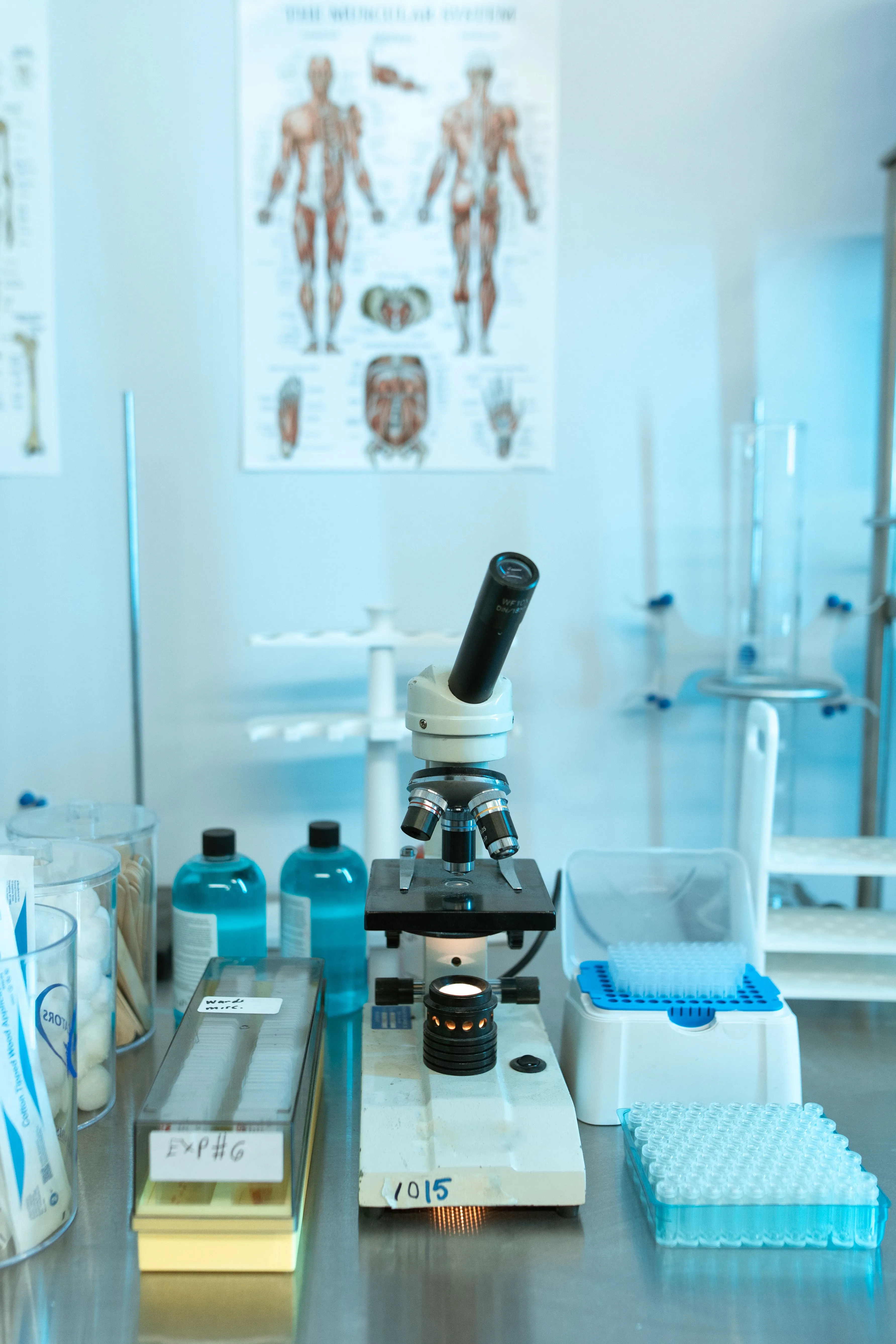
Reliable tissue analysis begins with the right instrumentation. Our imaging and staining platforms are validated for consistency across large sample sets, so results remain reproducible from the first slide to the last.
From brightfield and fluorescence whole-slide scanning to multispectral imaging and automated staining, each technology is matched to your markers, tissue, and study goals.
Whole-Slide Scanners
Brightfield and fluorescence scanning capture entire sections at high magnification for fully digital review.
Multispectral Imaging
Spectral unmixing separates overlapping fluorophores to enable high-plex marker panels without cross-talk.
Automated Staining Systems
Standardized, high-throughput staining platforms deliver consistent, reproducible results across large cohorts.
From Stained Section to Quantitative Data
Our analysis capabilities turn richly stained tissue into objective, reproducible measurements — capturing not just whether a biomarker is present, but how much, where, and in relation to which cells.
- Quantify biomarker expression by intensity, percent positivity, and H-score across whole sections.
- Map spatial phenotypes and cell-to-cell interactions within the tissue microenvironment.
- Perform single-cell segmentation and classification directly on tissue imagery.
- Assess marker colocalization and co-expression across multiplexed panels.
- Characterize the tumor microenvironment, including immune infiltration and stromal context.
- Preserve tissue architecture so molecular readouts remain anchored to morphology.
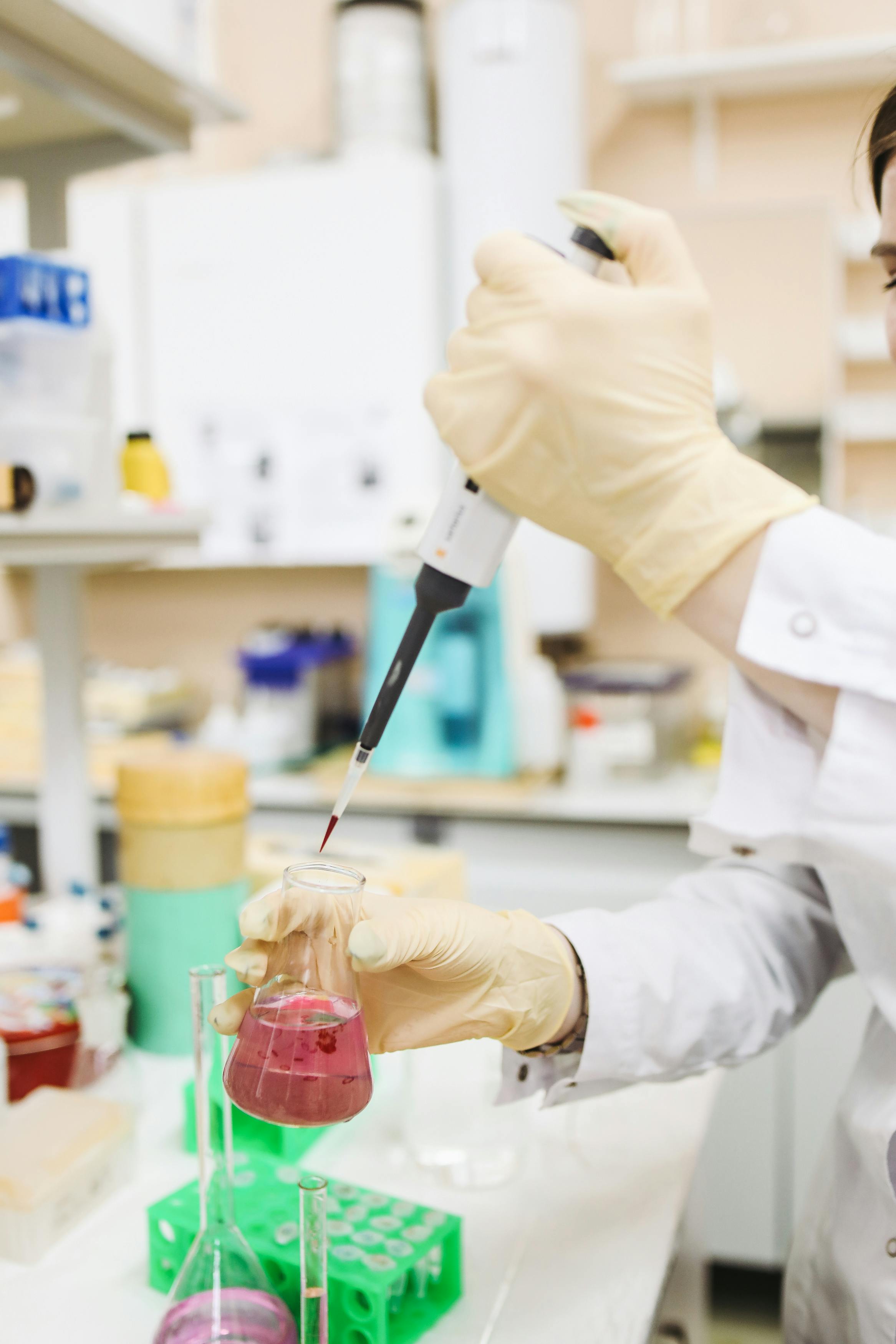
Digital Pathology & Image Analysis
Whole-slide imaging and algorithm-driven analysis deliver objective, reproducible results — with expert pathologists in the loop.
AI-Assisted Analysis
Machine-learning models accelerate tissue segmentation, cell detection, and region annotation with expert oversight.
Quantitative Scoring
Objective, reproducible quantification replaces manual estimation for consistent results across studies.
Spatial Analysis
Neighborhood and proximity analytics reveal how cell populations organize within their tissue context.
Pathologist Oversight
Board-certified pathologists review and validate every analysis to ensure accurate, defensible conclusions.
Applications Across Research & Clinical Programs
Tissue-level insight supports discovery, safety, and translational goals across a wide range of therapeutic areas.
Oncology Research
Characterize tumor morphology, biomarker expression, and heterogeneity directly within the tissue context.
Immuno-Oncology
Map immune infiltration and checkpoint marker expression to understand response within the microenvironment.
Neuroscience
Resolve marker expression across neural structures and cell populations in intact brain tissue.
Autoimmune & Inflammation
Visualize inflammatory infiltrates and tissue remodeling to understand disease activity in situ.
Biomarker Discovery
Localize and validate candidate tissue biomarkers with spatially resolved, quantitative readouts.
Drug Development
Support target validation and pharmacodynamic assessment with tissue-level evidence of activity.
Toxicologic Pathology
Evaluate tissue-level safety signals and morphologic changes throughout preclinical development.
Translational Research
Bridge molecular findings and tissue pathology to strengthen translational study designs.
A Guided Tissue Analysis Workflow
From sample accessioning to secure delivery, every step is tracked and quality-controlled.
Sample Accessioning
Tissue and blocks are logged, inspected, and tracked under controlled conditions on receipt.
Sectioning & Preparation
FFPE or frozen tissue is sectioned to precise thickness and mounted for downstream staining.
Staining & Labeling
Sections are stained via IHC, multiplex IF, ISH, or special stains on validated platforms.
Whole-Slide Scanning
Stained slides are digitized at high resolution into review-ready whole-slide images.
Image Quality Control
Each scan is checked for focus, staining quality, and completeness before analysis.
Image Analysis
Quantitative algorithms measure expression, positivity, and spatial distribution across the section.
Pathologist Review
Board-certified pathologists interpret and validate findings within the morphologic context.
Reporting & Delivery
Results, images, and datasets are compiled and delivered through secure, access-controlled channels.
Why Choose CNC Path Lab
A trusted partner for tissue imaging and analysis projects of every scale.
Expert Pathology Team
Board-certified pathologists and histotechnologists with deep experience in tissue-based analysis.
Advanced Imaging Platforms
Whole-slide scanners and multispectral systems validated for accuracy and reproducibility.
Quantitative Image Analysis
Objective, algorithm-driven scoring with expert oversight for consistent, defensible results.
Rigorous Quality Standards
Standardized protocols and multi-stage checkpoints safeguard data integrity at every step.
Secure Data Management
Confidential handling of tissue, images, and results across storage, transfer, and delivery.
Responsive Scientific Support
Guidance available from study design through execution and final reporting.
From Sample to Scientific Insight
Partner with a world-class precision diagnostics team
From biomarker discovery to AI-powered digital pathology, CNC Path Lab delivers rigorous, translational science that moves your program forward.
Life science innovators
come to CNC Path Lab
Ready to accelerate your clinical program? Our experts are here to help you design and execute the perfect laboratory strategy. Are you ready?