Immunohistochemistry
(IHC)
Protein-level biomarker detection in preserved tissue — specific, spatially resolved, and fully quantified.
Immunohistochemistry remains a cornerstone of tissue biomarker analysis and precision medicine. At CNC Path Lab, validated antibodies, automated staining, and AI-driven digital pathology turn a stained section into accurate, reproducible, decision-ready data.
Protein Expression in Native Tissue Context
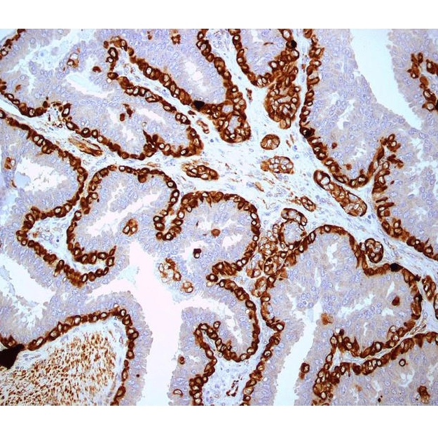
Immunohistochemistry (IHC) uses antibodies that bind specific protein antigens to reveal exactly where a target is expressed within a tissue section. A visible chromogenic or fluorescent signal marks each positive site, so protein presence, abundance, and localization can be assessed without disrupting the surrounding morphology.
Because IHC preserves tissue architecture, it links molecular information directly to histology — showing not only whether a biomarker is present, but in which cells and structures. That spatial context makes IHC indispensable across biomarker discovery, translational research, and clinical development, from early target evaluation through companion diagnostic support.
- Detect and localize protein biomarkers directly within intact tissue.
- Preserve morphology so results stay anchored to histology.
- Quantify expression objectively with digital image analysis.
- Scale from single markers to validated multiplex panels.
- Pair automated staining with expert pathologist review.
Comprehensive IHC Capabilities
A complete, quality-driven immunohistochemistry service spanning assay development, staining, digital pathology, and quantitative analysis.
Biomarker Detection & Validation
Localize and confirm protein targets in situ, with rigorously validated antibodies that deliver specific, reproducible staining across cohorts.
Chromogenic & Fluorescent IHC
Both brightfield chromogenic (DAB) and immunofluorescent detection, matched to the biology, sensitivity, and readout your study requires.
Manual & Automated Staining
Hand-optimized protocols for novel targets alongside fully automated platforms that ensure batch-to-batch consistency at scale.
Single-plex & Multiplex Assays
Classic single-marker staining through to multiplex panels that resolve several biomarkers on one section while preserving tissue.
Digital Pathology Integration
Every slide is scanned to a high-resolution whole-slide image, enabling remote review, annotation, and fully digital workflows.
Quantitative Image Analysis
Objective, software-driven scoring of intensity, positivity, and cell density replaces subjective estimation for defensible data.
High-Throughput Tissue Analysis
Tissue microarrays and streamlined pipelines process large sample sets efficiently without compromising staining quality.
Assay Optimization & Validation
Fit-for-purpose development, titration, and validation to establish accurate, robust assays aligned to your endpoints.
A Guided IHC Workflow
From sample receipt to final interpretation, every step is tracked, optimized, and quality-controlled.
Sample Receipt
FFPE blocks, slides, and tissue microarrays are logged, inspected, and tracked under controlled conditions on arrival.
Tissue Preparation
Sections are cut to precise thickness, mounted, and processed through deparaffinization and antigen retrieval.
Antibody Optimization
Antibodies are titrated and conditions tuned to achieve specific signal with minimal background for each target.
Staining
Validated chromogenic or fluorescent protocols are applied on manual or automated platforms for consistent labeling.
Digital Slide Scanning
Stained slides are digitized at high resolution into whole-slide images ready for analysis and archiving.
AI-Powered Image Analysis
Machine-learning models assist cell detection, tissue classification, and marker quantification under expert oversight.
Quantitative Reporting
Standardized scoring metrics and datasets are compiled into clear, reproducible, decision-ready reports.
Final Interpretation
Board-certified pathologists review and contextualize results to ensure accurate, biologically sound conclusions.
Automated Staining,
Digital Analysis
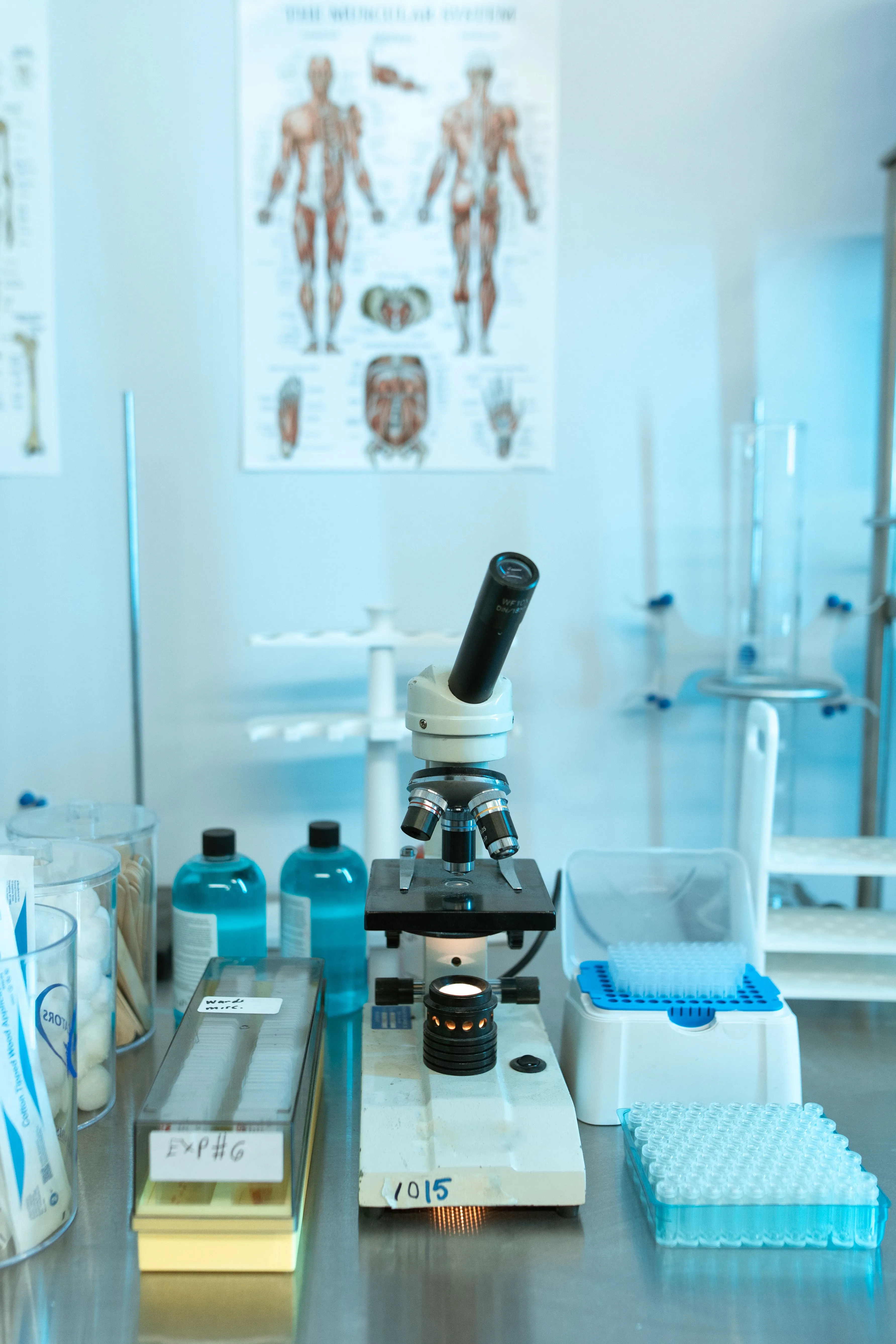
Reproducible IHC depends on the right instrumentation. Automated staining platforms standardize every run, whole-slide scanners digitize each section, and AI-assisted analysis converts stained tissue into objective, quantitative results.
Together, these technologies deliver consistent, defensible biomarker data across cohorts of any size — with expert pathology review keeping interpretation anchored to the biology.
Automated Staining Platforms
High-throughput autostainers deliver standardized, reproducible IHC across large slide volumes.
Whole-Slide Imaging
Digital scanners capture entire sections at high magnification for complete, review-ready images.
Digital Pathology
Fully digital workflows enable annotation, collaboration, and secure archiving of every case.
AI-Assisted Image Analysis
Deep-learning models accelerate segmentation, classification, and marker quantification.
Quantitative Pathology
Objective algorithms convert stained tissue into precise, reproducible numerical readouts.
Biomarker Scoring
Standardized scoring frameworks translate expression into consistent, comparable results.
Applications Across Research & Precision Medicine
Protein-level, spatially resolved insight supports discovery, translational, and clinical goals across a wide range of therapeutic areas.
Oncology Research
Characterize tumor biology, marker expression, and heterogeneity directly within intact tissue context.
Immuno-Oncology
Profile immune infiltration and checkpoint markers such as PD-L1 to inform response and resistance.
Biomarker Validation
Confirm candidate biomarkers with specific, spatially resolved protein-level evidence.
Drug Development
Demonstrate target engagement and pharmacodynamic effects across preclinical and clinical phases.
Translational Medicine
Bridge molecular findings and tissue pathology to strengthen study design and decision-making.
Companion Diagnostics
Support the development of tissue-based assays that guide patient selection and stratification.
Tissue Characterization
Define cell lineages, structures, and compartments to map the composition of complex tissue.
Precision Medicine
Provide spatially anchored evidence that supports individualized, biomarker-driven treatment strategy.
Validated Assays, Reproducible Data
Every IHC assay is optimized and validated for specificity, sensitivity, and reproducibility before it touches your study samples. Appropriate positive and negative controls, standardized scoring, and pathologist oversight ensure that results are accurate, comparable, and ready to support critical decisions.
- Antibody titration and specificity confirmation for each target.
- Standardized controls run alongside every batch.
- Objective, reproducible scoring via quantitative image analysis.
- Expert pathologist review of all final interpretations.
Why Choose CNC Path Lab
A trusted, end-to-end partner for immunohistochemistry and tissue-based biomarker programs of every scale.
Experienced Scientific Team
Scientists and board-certified pathologists with deep expertise in tissue-based biomarker assays.
High-Quality Laboratory Workflows
Standardized, validated protocols engineered for consistent, dependable results at any scale.
AI-Driven Digital Pathology
Advanced image analysis and machine learning for objective, quantitative interpretation.
Robust Quality Control
Multi-stage checkpoints and controls safeguard data integrity from receipt to final report.
Fast Turnaround Times
Efficient, well-orchestrated operations keep your program moving without sacrificing quality.
Flexible Research Support
Adaptable study designs and custom assays tailored to the biology and goals of your project.
End-to-End Biomarker Solutions
A single partner from assay development through digital analysis and expert interpretation.
From Sample to Scientific Insight
Partner with a world-class precision diagnostics team
From biomarker discovery to AI-powered digital pathology, CNC Path Lab delivers rigorous, translational science that moves your program forward.
Life science innovators
come to CNC Path Lab
Ready to accelerate your clinical program? Our experts are here to help you design and execute the perfect laboratory strategy. Are you ready?