Fluorescence In Situ Hybridization (FISH)
& In Situ Hybridization (ISH)
Advanced molecular pathology and genomic analysis for detecting DNA and RNA alterations directly within preserved tissue.
CNC Path Lab combines optimized probe based assays, high resolution imaging, and expert interpretation to identify gene amplifications, deletions, rearrangements, and fusion events at the single cell level while preserving the natural tissue architecture. This approach provides accurate spatial information and reliable molecular insights to support research and clinical decision making.
Genetic Insight, In Native Context
FISH and ISH detect DNA and RNA targets exactly where they occur — turning preserved tissue into precise, spatially resolved molecular data.
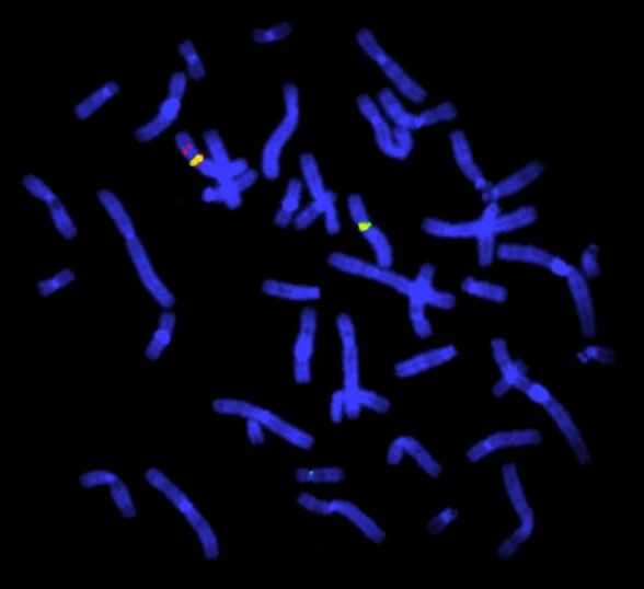
Fluorescence In Situ Hybridization (FISH) and In Situ Hybridization (ISH) are molecular cytogenetic techniques that use labeled nucleic acid probes to bind specific DNA or RNA sequences directly within intact cells and tissue sections, revealing genetic information exactly where it occurs in the specimen.
By hybridizing complementary probes to their targets, these assays pinpoint gene amplifications, deletions, rearrangements, and fusion transcripts without disrupting the surrounding morphology. Fluorescent or chromogenic detection then makes each signal visible under microscopy while preserving tissue architecture.
At CNC Path Lab, FISH and ISH underpin molecular pathology, biomarker analysis, oncology research, translational medicine, and precision diagnostics — linking genetic abnormalities to their spatial and cellular context so results remain both quantitative and clinically meaningful.
Why It Matters
- Single-cell detection of DNA and RNA targets within intact tissue.
- Genetic abnormalities identified without sacrificing morphology.
- Fluorescent and chromogenic signals for flexible visualization.
- Results anchored to tissue architecture for defensible interpretation.
Comprehensive FISH / ISH Capabilities
A complete in situ hybridization menu — from copy-number and rearrangement analysis to multiplex, quantitative, high-throughput workflows.
DNA & RNA In Situ Hybridization
Probe-based detection of both DNA loci and RNA transcripts directly within preserved cells and tissue sections.
Fluorescence & Chromogenic Detection
Flexible FISH and CISH readouts, from multi-color fluorescence to brightfield chromogenic signals reviewable alongside morphology.
Gene Amplification Analysis
Accurate quantification of gene copy-number gains such as HER2 and MYCN to inform biomarker and therapy decisions.
Gene Deletion Studies
Detection of losses and microdeletions, including tumor-suppressor and hemizygous deletions, with locus-specific probes.
Chromosomal Rearrangement Detection
Break-apart and dual-fusion strategies resolve translocations and structural rearrangements at single-cell resolution.
Fusion Gene Identification
Confident identification of clinically relevant fusions such as ALK, ROS1, and BCR-ABL1 driving oncogenic signaling.
Copy Number Variation Analysis
Objective, quantitative CNV assessment across target loci for stratification and biomarker characterization.
Biomarker Validation
Rigorously optimized and controlled assays that validate candidate biomarkers for research and clinical programs.
Companion Diagnostic Support
Fit-for-purpose FISH/ISH workflows that support companion diagnostic development and regulated study endpoints.
High-Throughput Tissue Analysis
Scalable processing of tissue microarrays and large cohorts with consistent, reproducible signal quality.
Multiplex Probe Assays
Multiple probes combined in a single assay to interrogate several loci or targets within one tissue section.
Quantitative Image Analysis
Whole-slide imaging paired with validated digital algorithms for objective signal enumeration and scoring.
A Guided FISH / ISH Workflow
From sample receipt to final reporting, every step is optimized, tracked, and quality-controlled.
Sample Receipt
FFPE blocks, slides, and cytology specimens are accessioned, inspected, and tracked under controlled conditions on arrival.
Tissue Preparation
Sections are cut to precise thickness, mounted, deparaffinized, and pretreated to expose target nucleic acids.
Probe Selection & Optimization
Locus-specific, break-apart, or fusion probes are selected and optimized for each target and specimen type.
Hybridization
Denatured probes hybridize to complementary DNA or RNA sequences under tightly controlled temperature and timing.
Washing & Signal Detection
Stringency washes remove unbound probe, then fluorescent or chromogenic detection renders each signal visible.
Whole-Slide Imaging
Slides are digitized across every relevant channel on high-resolution scanners for complete, reviewable images.
Digital Image Analysis
Validated algorithms enumerate signals, assess ratios, and quantify amplification, deletion, or rearrangement status.
Expert Interpretation
Experienced scientists and pathologists review every case against defined criteria and morphological context.
Final Reporting
Clear, quantitative reports with images and interpretation are delivered securely to support your decisions.
Applications Across Research & Precision Medicine
Genetically informed, spatially resolved insight supports discovery, translational, and clinical goals across therapeutic areas.
Oncology
Detect driver alterations and actionable biomarkers to guide research and therapeutic strategy across tumor types.
Hematologic Malignancies
Resolve translocations and copy-number changes central to leukemia and lymphoma classification and monitoring.
Solid Tumors
Characterize amplifications, deletions, and fusions in carcinomas, sarcomas, and other solid malignancies.
Companion Diagnostics
Support biomarker-driven therapy selection with validated, fit-for-purpose FISH and ISH assays.
Translational Research
Connect genomic findings to tissue pathology to strengthen study design and biomarker strategy.
Biomarker Validation
Confirm and quantify candidate biomarkers with spatially anchored, single-cell resolution.
Cytogenetics
Map structural and numerical chromosomal abnormalities directly within cells and tissue.
Precision Medicine
Enable patient stratification and targeted-therapy matching with genetically informed evidence.
Clinical Trials
Deliver standardized, reproducible molecular endpoints across multi-site and longitudinal studies.
Drug Development
Demonstrate target engagement and mechanism with tissue-level genomic readouts for pipeline programs.
Imaging & Analysis Technologies
Validated platforms that turn hybridized signals into objective, reproducible, and reviewable molecular data.
Fluorescence Microscopy
High-sensitivity multi-channel imaging resolves closely spaced FISH signals with clarity and precision.
Whole-Slide Imaging
Automated scanners digitize entire sections across every channel for fully reviewable, archival images.
Digital Pathology
End-to-end digital workflows enable remote review, collaboration, and secure long-term data management.
Automated Image Analysis
Validated algorithms enumerate signals and score ratios objectively for reproducible, defensible results.
AI-Assisted Interpretation
Machine-learning models accelerate detection and scoring under expert scientific supervision.
Multiplex Hybridization Platforms
Multi-probe assays interrogate several loci or targets simultaneously within a single tissue section.
Why Choose CNC Path Lab
A trusted partner for FISH and ISH programs of every scale — from biomarker discovery to clinical-grade molecular pathology.
Expert Molecular Pathology Team
Scientists and board-certified pathologists with deep experience in cytogenetics and in situ hybridization.
Advanced Laboratory Infrastructure
Validated hybridization, imaging, and digital pathology platforms engineered for consistency at scale.
High Analytical Accuracy
Optimized probes and stringent controls deliver sensitive, specific, and reproducible signal detection.
Robust Quality Control
Standardized protocols and multi-stage checkpoints safeguard data integrity from receipt to report.
AI-Powered Digital Pathology
Whole-slide imaging and algorithmic analysis add objectivity and speed to signal enumeration and scoring.
Fast Turnaround Times
Efficient, well-orchestrated workflows return high-quality results within timelines your program can rely on.
End-to-End Biomarker Solutions
From probe selection to reporting, we support the full biomarker journey under one roof.
Research & Clinical Support
Flexible services spanning discovery, translational research, and clinical-grade study requirements.
From Sample to Scientific Insight
Partner with a world-class precision diagnostics team
From biomarker discovery to AI-powered digital pathology, CNC Path Lab delivers rigorous, translational science that moves your program forward.
Life science innovators
come to CNC Path Lab
Ready to accelerate your clinical program? Our experts are here to help you design and execute the perfect laboratory strategy. Are you ready?